Glass half empty? We can’t see the glass anyway!

Robert profile picture

By Robert, who has aniridia

You have probably been asked this question before:

Are you a glass-half-full or a glass-half-empty person? 

Well, let me rephrase that for our community.

Are you a person living in a society that isn’t built for you?
Or are you a person that isn’t built for the society around you? 

As a rule, I would say we with aniridia are all people who were simply not built for the design of the world around us. Does that make us glass-half-empty? Well, probably yes—but none of us can see the damn glass anyway, so let’s not worry about it!

What I am trying to say is that we all spend our entire lives constantly adapting to this environment just to make life baseline manageable for ourselves. We have all held a piece of paper so close to our face that we fear we might snort it up our nose with our next breath, simply because we’d rather strain to read it than make a scene by demanding a larger font or asking someone to read it aloud. We do it for a quiet life. 

But you cannot be a ‘quiet life’ person and a change-maker at the same time. 

Let me tell you the truth about living in a world that wasn’t designed for us: we are clever, capable people who have immense value to offer. We possess an elite skill set that society is actively missing out on. Paradoxically, it is the world around us that is acting like the ‘glass half empty’ and choosing the quiet life. The system looks at us and says, “It’s too difficult, there aren’t enough of you to justify adapting, it takes too much time, and it costs too much money.”

Well, I am here to tell you that is about to change. 

Why? Because, if I am totally honest, I am a complete pain in the backside. But more to the point, I have the skills—and you have the skills—to shift the status quo. And no, you don’t have to be a pain in the backside yourself; you can leave that expertise entirely to me. Practise what you are good at, right?

Apart from not being able to see past the end of our noses, what do we have in common? On paper, possibly nothing. Our individual eye conditions are entirely unique to the bodies they come with; no two of us navigate exactly the same way. But do not despair: the fact that you have to focus on what is right in front of your nose is actually your greatest technical asset. It is the very thing that will change the world in our favor.

When you demand that the world changes to accommodate your journey, you inadvertently fix the world for absolutely everybody else. Your lived experience is an unmatched professional skill set. Everybody gets older. Everybody eventually becomes more physically or sensorily vulnerable. Because we have spent our entire lives adapting from birth, we are the natural experts in resilient design. It is time we used that skill set for the greater good.

Now, at this point, you do not have to don a cape or start wearing your underpants on the outside of your trousers like Superman. You don’t have to be a loud disruptor. Though, thinking about it, maybe I will try the underpants-on-the-outside trick next time—anything to get my point across to a local authority! 

You are probably thinking, “Dear God, Robert, get to the point.” 

Okay, I will. I am an Accessibility Consultant. What does that mean? I’ll tell you what it doesn’t mean: it doesn’t mean moaning about rigid regulations or helping corporations tick a box. It means showing society that we are worthy of being everyone’s absolute equal. It is not our job to clumsily adapt to an inaccessible environment; it is the environment’s job to adapt to us so we can navigate with dignity.

When I walk into a high street venue and the tap-and-go card machine inevitably fails, I am instantly stuck trying to read tiny buttons on a flat, highly reflective screen without a shred of assistance. What am I supposed to do? Smash my face against the terminal so close that I could tap my PIN number out with my tongue? Or am I forced to compromise my financial security and beg a stranger, “Please read this for me, here is my code”? 

I don’t want to choose between feeling that vulnerable or that anxious. What I want is simple: I want that business owner to have placed a low-cost visual magnifier right next to the card terminal. It doesn’t structurally redesign the hardware, but it gives me an immediate fighting chance at independent customer logic. Just imagine how much better the high street would be if every terminal featured that simple touch. That small change benefits the whole community—including the elderly lady whose sight is fading after a lifetime of perfect vision. She profits from that magnifier just as much as we do.

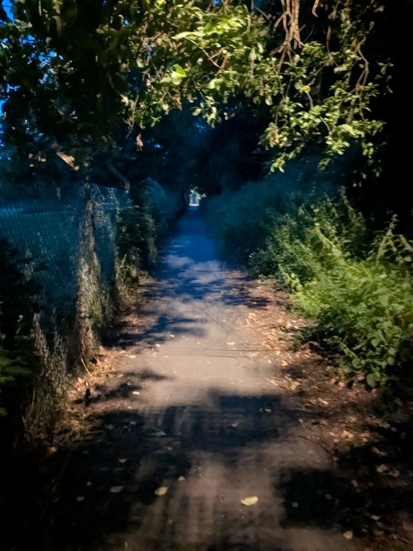
A dark path covered by trees and bushes

This is exactly why I am taking my lived-experience walkthroughs to the public realm. Right by my home in Wrexham, an old converted railway path serves as my primary walking route into town. Currently, it is a sensory hazard: unlit at night, aggressively overgrown with stinging nettles, and obstructed by dark green and black cycle chicanes that sit at ankle height—perfect for tripping over. 

Does this defect only threaten me? Absolutely not. Anyone can get stung; anyone can take an impact fall over an unpainted post in the dark. The difference is that the able-bodied public simply adapt and tolerate the decline because they don’t possess our specialized observation skills. 

Wrexham County Borough Council has chosen a bureaucratic roadblock over a simple fix, which is why I am putting on my metaphorical cape to highlight the issue. The solutions are blindingly simple and cost virtually nothing:

First, paint the cycle barriers a high-visibility, contrasting color instead of camouflaging them against the bushes. 

Second, establish a regular, logical maintenance routine. Don’t claim there’s no budget or staff; it takes one worker a few minutes during their regular bin-emptying route to clip back an intruding bramble or nettle before it causes an injury. 

Third, fix the lighting. Ingeniously, the council already has windmill-driven streetlights on this path. The only issue is that they haven’t installed enough of them. Placing them directly over the cycle chicanes to illuminate high-visibility safety paint solves the entire navigation risk.

Whether it is me navigating with limited sight or a neighbor walking home after a few too many drinks in town, we both possess the fundamental right to travel our local paths with confidence. 

Our lived journey insights can fix these broken layouts. To local authorities and high street businesses alike, I say this: give our community a moment of your time and invest in our professional expertise. Stop treating our autonomy as an afterthought, and see the untapped commercial and social value your disabled residents bring to the table. We aren’t here to make life difficult for everyone else; we are here to make infrastructure work better for humanity.

Now, if you will excuse me, I am off to remove my underpants from my head. Well, what did you expect? I can’t see a damn thing, so let’s not even talk about where the Superman cape ended up.

Posted in Patients' tales | Tagged , | Leave a comment

Be a leader of Aniridia Europe 2026-28

Aniridia Europe is recruiting volunteers to be directors, to lead and carry out its activities for the next 2 years.

Panel on stage with sponsors slide behind them

It’s an important and exciting role where you can influence the support provided to aniridia researchers, doctors, associations and patients, across Europe and beyond. Together the directors discuss issues and take decisions about what the organisation does. They also often do the tasks agreed, for example: liaising with doctors, running events and fundraising.

These activities take place mainly in English and online, so being comfortable with both of these is important. Directors need good critical thinking, administration and communications skills. As volunteers they need to have time, enthusiasm and dedication to the objectives of Aniridia Europe.

If this sounds like and interests you follow the process decribed at the form linked to below. If you would like to discuss or find out more about the expectations, browse the Aniridia Europe website and contact post@aniridia.eu 

Candidates for directors must be part of and nominated by one of Aniridia Europe’s full member aniridia associations such as Aniridia Network. Get in touch if with us if you are at all interested.

Posted in International | Tagged | Leave a comment

Parenting and aniridia: Online meet up

Saturday 20 June, 7:30pm via Google Meet – Sign up

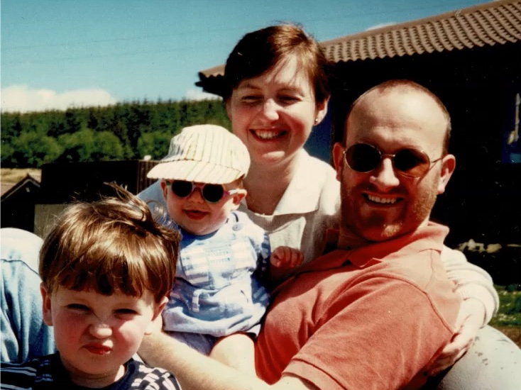
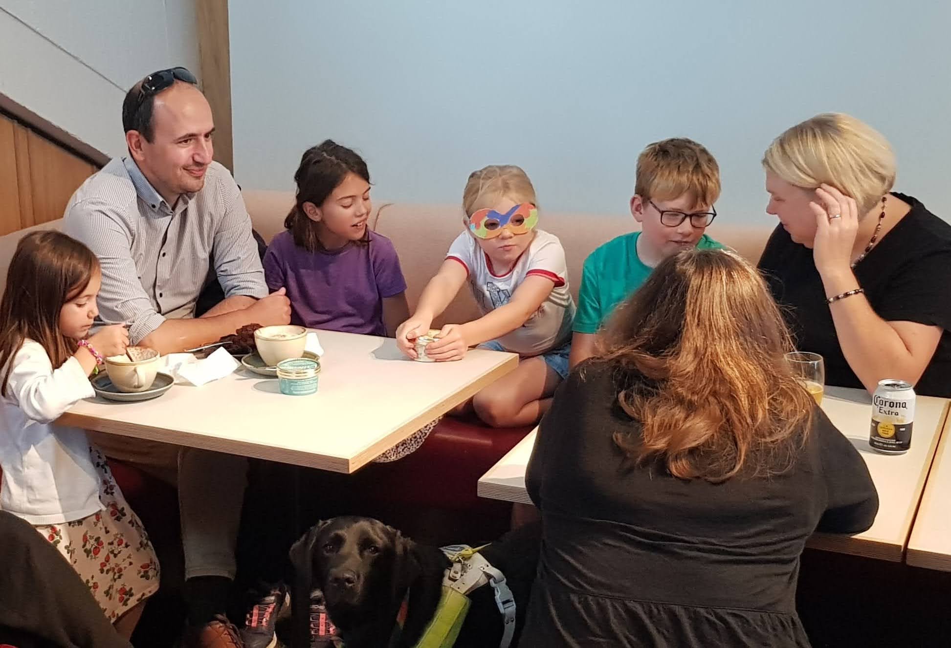
Dad and 3 children sitting at a cafe table

Learning your child has aniridia brings a range of thoughts and emotions. The effects on parents are not talked about enough, especially regarding men. Join us for an evening to mark Aniridia Day and explore parenting topics with 3 short talks and an open discussion for everyone.

Most of our parent members are mums – so this is a special invitation to the dads in our community – and for mums to get their partner to attend alongside them. And, patients, tell your parents to come. Join to hear fathers’ stories, and perhaps, share your experiences as a parent too. Everyone else is, of course, very welcome too.

What to expect

We’re delighted to welcome three speakers, each bringing a different perspective. Each talk will be about 10 minutes.

Harriet, Researcher

Harriet, an MSc Genetic Counselling student from Cardiff University, will be introducing a research project she’ll be carrying out with Aniridia Network. The study is looking at “What is the impact on family members of the diagnosis of aniridia in a child, and what would help?

Later this year Harriet will be recruiting parents and other family members for interviews about their experiences. She will explain what the project involves and how you can take part. This is a fantastic opportunity for you to directly shape research that could improve support for families.

Mark, sighted father: “Looking back on aniridia – 30 years of learning.”

Mark, 62, is the father of Harry, 29, who has aniridia. He will reflect on what he’s learned over almost three decades of supporting Harry — from diagnosis through childhood, school, and into adulthood — and the highs and lows along the way.

Mark’s full career was as an engineer in the Royal Navy followed by a consultancy. Now he is working part-time to allow more time for my hobby, supporting scouting.

Simon, aniridic father of aniridic son: “Empathising with my son – Raising a child with anirida when you have it too”

Simon, 43, will share what it’s like to navigate aniridia from both sides, as someone having it and bringing up a child with it. His son is going through many of the same things he did growing up. Yet the world has also changed and mnay of the challenges new or differnet.

Simon has aniridia, nystagmus, optic nerve hypoplasia and foveal hypoplasia. He is married with one child and works as an IT engineer and have worked

Conversation

After hearing from our speakers, we’ll open up for a group discussion. We hope Mark and Simon’s stories will encourage you, especially dads, to share your fears, approaches and recollections of parenthood, and family life with aniridia. There’s no pressure to speak; you are welcome to simply listen, as well as join in with questions, comments, and stories of your own.

If possible the three talks will be recorded and shared on our YouTube channel afterwards. The discussion afterwards will not be recorded to enable open conversation in private.

Why now?

Aniridia Day logo

There are 3 important adjacent dates that we want to celebrate together, while leaving space for family gatherings on the Sunday:

How to join

We’d love to see as many of you there as possible to mark Aniridia Day. Sign up to get the link to join us on Saturday evening.

If you know of a parent or carer, particularly a man, who would benefit from attending, please encourage them to sign up.

We are looking forward to a warm, honest, and hopefully inspiring evening together.

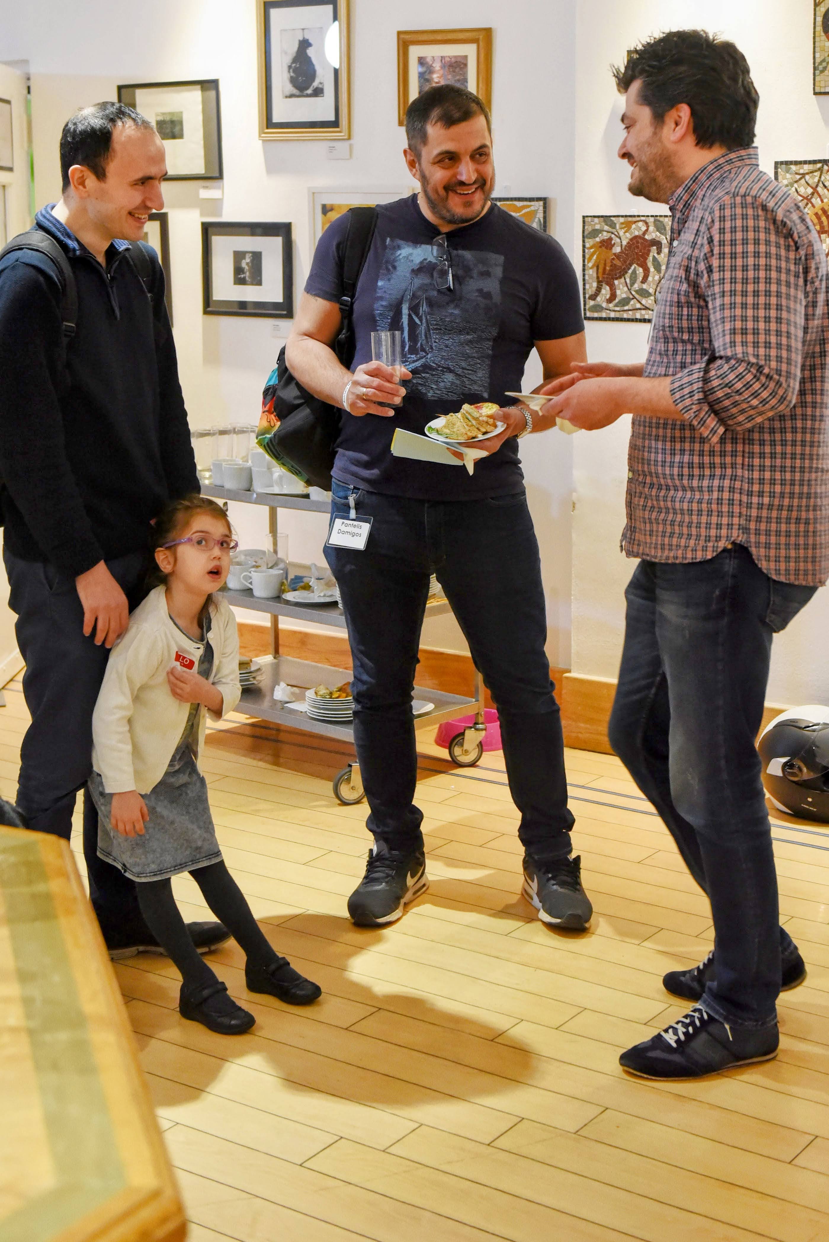
3 dads standing talking to each other while eating lunch at a conference. Small girl leaning against one of them.
Posted in Parents' accounts | Tagged , , | Leave a comment

Attending the 8th European Aniridia Conference as a professional

James, Nicky and Katie in front of European Aniridia Conference banner
Nicky with Aniridia Network trustees James and Katie

Thanks to the generosity of Aniridia Network, I was fortunate enough to attend the 8th European Aniridia Conference. This three-day event brought together international clinicians, researchers and families to discuss all aspects of aniridia.

I am an ophthalmology registrar and PhD student to Professor Mariya Moosajee at UCL and Moorfields Eye Hospital.

I sucessfully applied to Aniridia Network to pay my expenses to attend the event in Sofia, Bulgaria.

The conference by Aniridia Europe covered not only the latest research being done in the field, but also aspects of clinical management and how these should be refined for different patients.

In a refreshing step away from the norm for conferences, we heard from patients about their experiences and what would be most beneficial for them.

Particularly interesting for myself, as a clinician and researcher, was hearing about some of the cutting-edge research that is being done with stem cells and targeted therapies to develop new treatments for aniridia-associated keratopathy. This included both repurposing existing drugs as eye drops and creating new types of drops, both of which show promising results and offer hope for the future. Research into stem cells grown from skin samples from aniridia patients is expanding our knowledge of the biological changes that underlie aniridia-associated keratopathy and offers more avenues for this too.

The best aspect of this fantastic conference was the opportunity to connect with other researchers from around the world, discuss the work we are doing and bounce around ideas to refine the focus and direction of our projects. I have come away from this with information that will benefit both my current research and also many ideas for future studies and collaborations.

I met Aniridia Network trustees James and Katie at the event too. Thank you again, Aniridia Network, for providing me with this amazing opportunity. I have left the conference with not only new friends, colleagues and information, but also a resolution to give more time and thought to my patients, to ensure I am providing individualised support where and when it is needed.

Katie and Nicky together
Posted in International, Medical staff talking | Tagged , , , | Leave a comment

Sudden sight loss on Sunday morning

12.10am Sunday, Home: What’s gone wrong?

I was reading before going to bed when the text suddenly went blurry. “That’s very worrying,” I said out loud. 

Within seconds, my right eye was only aware of the screen brightness. There was  a greasy film with black dots floating around on it. My left eye was unaffected. A sense of dread descended and my pulse quickened.

Very blurry image simulating the view with a cloudy vitreous. No text can be discerned and people are only vague shapes

I was not totally unprepared. Since birth I have had a visual impairment called aniridia. I’ve always been short-sighted, my acuity is 6/36 (typical sight is 6 times better than mine). Further sight loss was also likely at some point, not just due to old age. 9 years ago cataracts developed over a few months in both my eyes. I went from having no aids to needing a white cane and text read out to me. Operations restored my sight to the (low) level it had been previously. Compared to others with the same condition, I’d been relatively lucky with my acuity and its stability. So I have a good enough understanding of eye anatomy and my risk factors to take this seriously.

All that said, it was very scary; one eye had instantly returned to the worst period of the cataracts. 

But I stayed calm and practical.

I tried my regular lubricating eye drops first, in case something was simply sitting on the surface of the eye. They made no difference. 

I turned to an AI assistant. I described the above medical history and what had happened, asking it to respond as an expert ophthalmologist. The response was measured and clear. It outlined several possible causes, among them what I had guessed: retinal detachment. It advised that I should see a doctor immediately. 

“What does that mean in the middle of the night at the weekend?!”

Fortunately, I live in London and have regular checkups at the world-renowned Moorfields Eye Hospital. I knew they had a 24-hour accident and emergency department. I looked up the details. I figured that the sooner any (waiting for) treatment could begin, the better, and it was likely to be quieter now than in the daytime.

I went downstairs, where my wife was watching TV. I told her what had happened and that I thought I needed to go to Moorfields right away. Equally concerned she agreed. I said there was little use her coming to wait-up with me during the early hours: she should go to bed and I’d call if and when I needed help. 

1:00 Sunday: Into the dark

We prepared a bag with things to keep me going: food, reading material and an inflatable pillow!

We hugged with worry and trepidation before I set off. I took an Uber into town, passing by oblivious revellers in Stoke Newington and Hackney. 

1:21am Sunday, Old Street: Unexpected challenges

The emergency department was quieter than I had expected, mostly empty with lights off. The receptionist took my details and went to speak with a clinician. She returned quickly with instructions I had not anticipated.

My symptoms pointed to an issue that was not absolutely critical. It could be investigated when the department was fully staffed. I should come back at 8am. I was given a chit to bypass triage at least.

It was now nearly 2am. I faced the prospect of getting home, sleeping and returning within six hours. I walked to 4 nearby hotels but found them all fully booked. I considered going back to snooze in a hospital chair. But I feared I’d be sitting there for many more hours later in the day too.

I made my way home and got into bed around 3.15am.

8.00am Sunday: Moorfields Eye Hospital Emergency Department

After 2½  hours of sleep and another train ride I was back at the hospital, along with many other worried people in something like the same condition as mine.

At 9.00am a nurse did the standard sight tests. My right eye could only discern the vague shape of a hand moving in front of a light. 

At 11.00am I saw a doctor who did an ultrasound examination. While he could see blood in the liquid vitreous that fills the eyeball, he could not see a retinal detachment, though that had to be suspected. He referred me to the Vitreoretinal Emergency Clinic. He explained that they do examinations in the morning to prioritise their surgery in the afternoon. So if I went up right away I risked not getting dealt with today. It was best to go first thing the next day.

On my way home I noticed that I was bumping into things as I couldn’t judge distances, and was unaware of people close by on my right.  

Exhausted, after lunch, I slept for the rest of the day.

8.00am Monday: Moorfields Vitreoretinal Emergency Clinic

As ever proceedings began with an acuity test. I was just about able to see a large letter held about a meter away from my right eye.

Another ultrasound again could not clearly identify the cause of the vitreous clouding, nor therefore the likely prognosis. 

The doctor explained my choice.

Option 1: Watchful waiting

Do nothing and hope that over a few months the blood and cells floating in the vitreous will settle and get reabsorbed. The advantage was that there would be no risky intervention on an eye already complex due to aniridia. The disadvantage was that there was no guarantee it would clear, as the cause could continue. More seriously, if there was an underlying retinal issue, it would remain undetected and could deteriorate further without any clear warning signs. I’d have to visit the hospital regularly for checks.

Option 2: Surgery

A vitrectomy would remove the cloudy vitreous fluid and enable the surgeon to examine the retina to find, and if necessary, treat the problem. In time the eye would naturally refill it with clear fluid, restoring sight. The recovery would likely be shorter: weeks, with only one follow up visit. On the other hand, on top of the standard surgery risks, for people with aniridia, there is an elevated risk of inducing glaucoma or even aniridia fibrosis syndrome – which could reduce sight in the longer term.

Decision

I chose surgery. My level of sight was already very low. I hoped for the faster restoration and to avoid the uncertainty of both the cause being a one-off or ongoing and whether it would be improving or worsening over time. That it was even offered reassured me that surgery was worthwhile. That was my personal calculation, and it will not be the right one for everyone. 

10.00am Monday, London: Me, myself and eye

Surgery was not until the afternoon, so I had 3 hours to fill. I tried to visit one of the nearby museums, only to discover that they were all shut on Mondays! I eventually went to UCL where the preserved body of philosopher Jeremy Bentham is on display. After the treat of a burrito for lunch, I made my way back to the hospital.

1.00pm Monday, Operating Theatre: Making light of it

I was actually first on the list for surgery.

It was performed under local anaesthetic. Having previously had cataract operations under general anaesthetic, I was apprehensive about this. I did not relish the idea of seeing someone come at my eye with a scalpel. In practice, the worst part was the preparation: the anaesthetic injections and doing whatever they do to keep the eye open. I try not to think too much about that or the operation itself, let alone Google it!

Once underway, there was no discomfort. My left eye was shut/under a plastic cover. My right eye was too clouded to make out what was happening.

What I could perceive was both strange and fascinating: the silhouette of a long thin device akin to a vacuum’s crevice attachment. I watched it suck out the cloudy vitreous and black floaters. Next it injected a dye (akin to seeing food colouring being added to water) to help the surgeon identify bits that still need to be removed. Then it hoovered up the dye again. At other times it was a bit like looking through a kaleidoscope. 

There was a trainee observing and asking questions. So I heard the surgeon explaining what they were doing. I already knew or had just read up enough to follow along and get the idea that it was positive. 

They found a vasoproliferative lesion (abnormal blood vessels on the retina) which had bled. They chose to leave it alone rather than risk fiddling with it. Critically, there was no retinal detachment. They refilled the eyeball temporarily with saline and an air bubble (not a longer-lasting gas). There was also no need to maintain a specific posture (such as face down) during weeks of recovery. On the spectrum of possible causes and outcomes, this was all at the less serious end.

James with a dressing taped over his eye

The procedure had taken approximately twenty minutes. 

Soon, they were sealing the incisions and taping on a patch. I sat up on the operating table to get into a wheelchair and be taken to recovery.

I was given a cup of tea and custard cream biscuits – oh, and medication for the coming weeks. I was declared unfit for work for 2 weeks. Within an hour I was free to go. I walked out, got in a taxi and was home by 4pm.

Tuesday, Home: Not a patch on normality

As instructed, I removed the eyepatch and cleaned the eye with cooled boiled water. It was bloodshot and sore. I took paracetamol for the aching. I had two types of eye drops to take 4 times a day to reduce inflammation and prevent infection. 

Sight was still extremely limited, similar to before the operation. What was different was the rough black line across the middle of my vision. This was the boundary between the air bubble and saline. As I shook my head I could see it sloshing around. As advised, over the next few days the bubble got smaller as the air dissipated to be replaced by natural fluid. 

Very blurry image simulating the view post surgery. A black horizontal line is half way up the image. No text can be discerned and people are only vague shapes
James wearing a grey eyepatch

Counterintuitively, the line descended rather than rose in my field of view. I tried in vain to understand why. I believe this paper explains it: Optical Effects of Intraocular Gas Tamponade.

An AI assistant explained it in this simplified way:

“The lens inverts the image projected onto the retina — light from above hits the lower retina, light from below hits the upper retina. As the air bubble shrank, its lower boundary rose inside the eye, unblocking the upper retina first. Since the brain corrects for the lens inversion, this appears as the line moving downward in perceived vision.”

The jiggling line was quite distracting; it made me feel a bit motion sick. The blurriness interfered with the sight of my left eye. I got a patch to reduce the effect of both.  

Thursday, Home: Keeping my spirits up

3 days later, the air bubble formed an arc when looking forward and a circle when looking at the floor. I could jiggle it around. It continued getting smaller. It was like looking through a spirit level. If other surgical gases had been necessary the bubble would have lasted weeks.

Blurry image of Moorfields entrance, simulating the view with a reduced bubble. The black line is now a semi-circle anchored on the bottom corners of the image. Text can be discerned and potentially read if large or backlit. Major features of people such as hair and clothes can be distinguished

Meanwhile my vision slowly began to get clearer. While it lasted, the bubble actually was the best area and even had a slightly magnifying effect. 

I found brightness difficult as it was scattered by the blurriness to become glare. I could see backlit screens, but white backgrounds overwhelmed black text. 

I found that switching my computer and phone to dark mode (white text on a dark background) helped significantly. Very encouraging.

Friday, Home: Words of encouragement

4 days after surgery, my right eye could read text on my phone again. It was difficult, like looking through a condensation-covered window, but readable. That felt wonderful.

Slightly blurry photo of the front of Moorfields Eye Hospital. Text can be read and people's features seen. No black edge of a bubble.

Tuesday, Vitreoretinal Emergency Clinic: Bubble busted

Over the weekend the bubble had shrunk to be a circle, only visible with my neck bent to look at the floor. It then disappeared.  

However, I was experiencing really bad headaches/pain. I had thought they were due to the surgery or disorientation. But that had passed while the ache around my right eye had grown. It kept me awake at night and incapacitated me the following day – though paracetamol helped a lot.

After phoning for advice, I returned to the Vitreoretinal Emergency Clinic. 

As I suspected, the pressure in my right eye was high (30 mmHg), compared to my left eye (20 mmHg) and my normal readings.

My medication was changed: 

  • fewer dexamethasone drops (anti-inflammatory steroids) that can cause pressure to rise. 
  • new anti-inflammatory and pain relief, non-steroidal drops (Acular – Ketorolac Trometamol) 
  • drops to reduce the production of fluid in the eye to reduce the pressure (Cosopt/Codimaz – Dorzolamide/Timolol).

Sunday: Working well

Two weeks after the bleed and surgery, my vision is almost back to normal (for me) -I think. The main difference between the vision in each eye is that the right has a yellow tint. It is more sensitive to bright light but I think it always was the one I’d squint most anyway. Hopefully, the tint will reduce as the fluid clears further.

I’m due to return to work. I have a follow-up appointment at Moorfields in a few days.

Reflections

One thing I found difficult during recovery was the lack of illustrations of the experience to compare to my own. That’s partly why I wrote this post.

One account suggested that the bubble area should be relatively clear. I was worried at first that was not the case for me.

The sudden loss of functional vision is frightening. I tried to take each step at a time, noting that I still had a half-decent eye and that I had a lot of great support around me, at home, at work and in hospital to deal with the problem however it turned out.

The care I received at Moorfields was excellent and amazingly fast. From the moment the bleed occurred, until surgery was about 37 hours. Then nearly completely vision restoration within a fortnight.

I feel some things should be better though. They have not had an impact on me but I can see how they could on other people.

  1. When I looked it up, the emergency phone line for Moorfields A&E was not 24 hours or even 7 days a week. One call would have avoided my early hours dash to central London. Perhaps it could have diverted to NHS 111.
  2. The hospital could have offered me a comfortable place to sleep between 2am and 7am rather than sending me away.
  3. I’d like the medical staff to indicate they have looked at my extensive eye health notes before speaking to me. Because I couldn’t be sure they had all the information, I felt I needed to explain facts such as my underlying aniridia and prior cataract operations. It was unnerving.

Thank you

I want to thank everyone at Moorfields Eye Hospital who was involved in my care. I am grateful also to my regular ophthalmologists, who were supportive when I informed them by email. 

Most of all, I want to thank my wife. She sent me off into the night with composure and kindness when it was very concerning for her too. She has been wonderful and patient with the patient throughout.

If similar has happened to you

Sudden vision loss needs to be assessed urgently. Get in front of an eye doctor as quickly as you can.

Consider the options of waiting and surgery in advance if you can. If you are facing the same decision, talk it through honestly with your surgeon. There is no universally right answer. The choice depends on your eyes, thoughts on surgery, circumstances, and what you can live with. 

For those with aniridia or other pre-existing conditions, the additional surgical risks are worth discussing specifically, not dismissing.

My very best wishes for a swift and successful recovery to you.

Posted in Patients' tales | Tagged , , , , , | 3 Comments

Meet-ups for Rare Disease Day 2026

Group of mixed aged people at a cafe table looking at the camera

We held 4 friendly online/in-person gatherings of patients and their relatives to celebrate Rare Disease Day 2026.

  • London Euston Station: Starbucks: Aaron, Johnathan, Keith, James, Emily and children
  • Cambridge: M&S Food Cafe, Market Hill: Tony, Andy, Laura, Anastasia and children
  • Manchester: Piccadilly Tavern: Gemma and Katie
  • Online via Google Meet: Annie, Cerys, Simon, Johnathan, Andy, James

It can be hard having a rare disease and feeling like you are navigating this journey alone. So we enjoy talking to ask questions and hear from people who can share insights into living with aniridia. It makes a difference and empowers us.

To fit with the theme of Rare Disease Day, we focused on equity in medical care, discussing positive and negative experiences as well as self-advocacy with doctors.

2 children, 1 woman, 2 men eating and talking at a coffee shop table that has a Aniridia Network poster and white cane on it.

If you would like to host a meet-up in your area, email meetup@aniridia.org.uk!

Rare Disease Day

Rare Disease Day logo

We host events around this time of year to mark Rare Disease Day. It’s an international celebration held annually on the 28 February (29th on rare occasions!)

It’s aim is to work towards equity in social opportunity, healthcare, and access to diagnosis and therapies for people living with a rare disease.

With over 300 million people worldwide affected by rare diseases, this day serves as a crucial platform to highlight the importance of research, support, and resources for those who often feel overlooked. It’s a day to stand in solidarity, share stories, and foster a sense of community among those facing unique challenges.

Since its creation in 2008, Rare Disease Day has played a critical part in building a community that is multi-disease, global, and diverse– but united in purpose.

Find out more and get involved in supporting Rare Disease Day

Calling for equity

Equity for people living with rare conditions is equitable access to diagnosis, treatment, health, social care and opportunity

The theme of Rare Disease Day in the UK is ‘equity’, defined as ‘meeting people’s specific needs and eliminating barriers preventing their full participation in society.’

People living with a rare condition face many challenges with accessing health and social care services. This can happen for many reasons, including a lack of knowledge among healthcare professionals to support timely diagnosis and appropriate treatment, or limited access to specialist centres where care is provided.

People with a rare condition may have worse health outcomes than people in the general population due to the limited services and support available to them, and the support for people with different rare conditions is highly variable. Differences in health opportunities and outcomes which are systematic, avoidable and unfair are defined as health inequities, which are important to address to ensure that services are equitable for all people in the UK. (England 2025 Rare
Disease Action Plan – summary of health inequity scoping review
).

Why your voice matters

People living with, or affected by, a rare condition are the experts in their rare conditions. Your personal story is the most powerful tool for driving change. Sharing your experience is important because it can help to: – Humanise statistics and data: It shows decision-makers and the public the reallife impact of rare conditions. – Inform better policy: Your lived experience highlights the gaps in the system, such as diagnostic delays or lack of coordinated care, providing evidence for the changes we campaign for. – Build community: It connects you with others, reducing isolation and fostering a strong, united community. By raising your voice this Rare Disease Day, you contribute directly to the ongoing work to implement the UK Rare Diseases
Framework
and secure a future where everyone with a rare condition has equitable access to the best care.

Key messages

When raising awareness and campaigning, use a few clear, impactful messages that tell people and policymakers what changes you want to see and why. It is important that your messages reflect what matters most to you, but you can give your messages greater weight by connecting them to the shared concerns of the 3.5 million people in the UK living with a rare condition. Here are some examples

  • Call for equity for rare conditions
    Everyone with a rare condition deserves fair and equitable care from the NHS, no matter how rare their condition is. Equitable care means addressing individual needs, not treating everyone the same.
    Read a briefing on equity for rare conditions from EURORDIS
  • Renew the UK Rare Diseases Framework
    The UK Rare Diseases Framework must be renewed, refreshed, and underpinned by ring-fenced funding to drive continued progress beyond 2026.
    Read about our campaign for a renewed UK Rare Diseases Framework
  • Improve care coordination
    Only 1 in 10 adults in the UK living with a rare condition have a care coordinator to help organise different aspects of their care. People with rare conditions need well-coordinated, holistic care pathways and access to care coordinators.
    care coordinators.
    Read a factsheet on coordination of care for more information
  • Increase healthcare professionals awareness of rare conditions
    Healthcare professionals need increased awareness and training of rare conditions to prevent misdiagnosis and improve early support. Medics For Rare Disease (M4RD) provide information and learning resources to healthcare professionals who want to know more about rare conditions.

Posted in Aniridia Network news | Tagged , , | Leave a comment

Bursary for UK professionals to attend European Aniridia Conference 2026

The panel of experts seated on the stage

Aniridia Network is offering to fund UK professionals to take part in this year’s European Aniridia Conference (EAC), 17-19 April in Sofia, Bulgaria.

EAC enables the sharing of scientific knowledge about the rare genetic eye condition aniridia. Its goal is to prevent sight loss and deal with aniridia’s effects. It brings together patients and the world’s top experts to upskill the clinical, research and aniridic communities.

We are keen for doctors and researchers based in the UK & Ireland to take part, to get insights from those developing and delivering treatments and people living with this complex visual impairment.

We want to improve medical treatment and scientific knowledge through:

  • enabling networking
  • encouraging collaboration.
  • accelerating progress
  • avoiding duplication

If you are unable to get support from your institution; our charity will consider paying for your conference ticket plus reasonable travel and accommodation expenses. We would especially like to help people early in their careers.

Apply

Write up to 250 words to info@aniridia.org.uk explaining:

  • why you need the bursary
  • how it will meet the goals above and ultimately, (potentially) help people affected by aniridia
  • specifics about your work/projects in a way understandable by patients and their relatives.

You can also include (a link to) your resume for background.

If you attend the conference, we expect you to write a blog post for our website and/or speak at our next event, about your experience and what you got from it.

Please pass on details of this opportunity on to people who may be interested.

Posted in Aniridia Network news, Research | Tagged , | Leave a comment

Management of glaucoma associated with aniridia

John Brookes

Talk by Mr John Brookes, Moorfields Eye Hospital, at Conference 2025

A presentation about aniridia issues, particularly regarding how glaucoma is treated in children and adults with aniridia, from medical to laser and surgery.

Mr Brookes trained in London and qualified in 1993, subsequently specialising in ophthalmology and further, in paediatric glaucoma, for which he has been a consultant at Moorfields Eye Hospital since 2004. His main interest is in secondary glaucomas in children, such as aniridia and their surgical management.

Transcript

[James] Fantastic to have with us now John Brookes, who is also one of our medical advisors for Aniridia Network, recently appointed. Take it away John, thank you very much.

[John] Thank you very much. So thank you for the very kind invitation to come and talk to you today.

So just a bit about me, I’m a consultant at Moorfields Eye Hospital, which I have been for about 22 years, and I also work at the Royal Victoria Hospital in Belfast, and so I spend a day a week in Belfast seeing all the children from Northern Ireland with glaucoma. So I deal with the whole range of paediatric glaucoma. So I’m going to talk a little bit about what my experience is in treating children with aniridic glaucoma.

So as you know aniridia is quite a rare condition. It was first described by somebody called Barratta in 1818. I’ve tried to find out who Barratta is, but have been unsuccessful, but he described it quite a long time ago.

It’s a bilateral condition, as you know, so it affects both eyes. And it’s a pan-ophthalmic disorder, so it’s not only the iris that is involved in the condition. And obviously I’ll show the other clinical effects that aniridia can cause in children.

It’s rare, so it’s estimated to affect between one in 40 to one in 100,000 children. And I’m going to really concentrate this talk on dealing with children, which is what I deal with on a daily basis.

You can see on the clinical photographs on the left, total aniridia, and obviously there are variable degrees of iris hypoplasia. So hypoplasia is an underdevelopment of the iris. And these two photographs show total aniridia. So there’s a complete absence of the iris.

And on the upper slide, you can see these little bobbles here, which are called the ciliary processes, and we’ll come back to those. These are the parts of the eye that manufacture aqueous. This is the fluid that fills the front part of the eye. And that’s very important when we’re talking about raised intraocular pressure, which is what we need to deal with when we’re talking about glaucoma.

I’m sure you’ll have heard about this from Professor Moosajee earlier on this afternoon, that about 85% of cases are familial. So these are autosomal dominant cases. So that means that you only need to inherit one faulty gene from a parent to exhibit the disease itself.

About 15% of cases are thought to be sporadic, so there’s no family history. And it’s important because, as you know, these can be associated with other systemic problems, such as Wilms tumour.

You can see on the lower slide here, this is a case of aniridia, but only partial aniridia. So the absence of the iris is only in the upper part of the eye there. And again, this top slide’s total aniridia. And you can see the ciliary processes, which are the cells that produce the aqueous humour. The incidence of aniridia is the same in boys and girls.

Obviously, photosensitivity is a very important symptom of aniridia. And you can see this child has to have very dark glasses to cope with his photophobia. And we couldn’t really take them off him to have a look at his eyes until he was under general anaesthetic.

But there’s a variety of eye conditions that can stem from aniridia. Nystagmus is an involuntary jerking movement of the eye that happens in children with poor vision from very early on.

Squint is a non-alignment of the eyes, so one eye turns inwards or outwards. That’s usually because of a condition called amblyopia, otherwise a lazy eye. Because children, as they’re developing their vision, often in medical conditions, often have one eye that’s a little bit stronger and sees better than the fellow eye. And if that happens, then the fellow eye, the brain connections don’t develop normally. So they ignore all the vision from the poorer eye. And that’s called a lazy eye or amblyopia. And we have to patch the good eye, the better-seeing eye, to force the fellow eye to start seeing a bit better, to strengthen it up.

And these conditions, squints, amblyopia, refractive errors, are very common in aniridia.

One of the very big problems for vision reasons in aniridia is corneal pannus. This is a progressive fibrovascular scarring around the cornea that affects the peripheral cornea that can spread more centrally.

This is due to the absence of what we call limbal stem cells. So these are specialised cells that develop into corneal epithelial cells. And there’s an absence of this in aniridia. So instead of the clear cornea regenerating itself, the scar tissue replaces it and that can have a very big effect on vision.

It’s also very difficult to treat this, because if it gets to a stage where it needs a corneal transplant, for example, because of the vascularisation of the abnormal blood vessels, that can make the rejection of a new cornea much more likely. So corneal transplants often fail, I’m afraid, in aniridia because of this problem.

Cataracts are very common in aniridia. They account for about 85% cases of aniridia. And we often have to carry out cataract surgery to improve people’s vision.

Optic nerve and foveal hypoplasia, and certainly foveal hypoplasia is almost universal in aniridia. And this is an underdevelopment of the fovea, which is the central part of the retina, which deals with the very fine focusing. And this is the reason why children in infancy have poor vision. Poor vision happens in aniridia in older age because of either cataract, corneal problems or glaucoma.

And this is obviously my main focus. And about 50% of children with aniridia may well develop glaucoma. And it’s probably one of the most important aspects of aniridia to talk about, because this is the condition that is irreversible. Other conditions within aniridia can be treated. And glaucoma can certainly be treated and stabilised, but any damage that happens from glaucoma is irreversible.

A couple of syndromes in aniridia you may well have heard of. WAGR, that occurs because of a deletion on a particular chromosome – chromosome 11. And it’s thought to account for about 30% of sporadic cases of aniridia.

And this is the importance of referring children to paediatricians because we need to screen for Wilms tumour, and do the genetic testing that Professor Moosajee would have talked about earlier.

Gillespie syndrome is quite a rare syndrome associated with aniridia, and this accounts for about 2% of cases of aniridia. And this is an autosomal recessive disease. And so both parents need to be carriers for an affected child to develop Gillespie syndrome. And so one faulty gene needs to be acquired from each parent. But importantly, this is not associated with Wilms tumour.

So I normally go through talks and go through consultations and see children for 10/15 years. And I think the parents and the children know everything about glaucoma. And then somebody will say “What exactly is glaucoma?” So I thought we’d just address that issue first.

It’s an interesting definition, really, because at the end of the day, glaucoma is a disease of the optic nerve. And if you’re in glaucoma clinics, you’ll be very obsessed by intraocular pressure. But in fact, you don’t need to have a high intraocular pressure to have glaucoma.

So glaucoma is an optic neuropathy. So it’s a disease that affects the optic nerve. Now having said that, all children’s types of glaucoma – aniridia, and all other types of paediatric glaucoma – only occur when a child has a high intraocular pressure. And we’ll talk about why that might happen in a minute.

Now, adults can have glaucoma even with a normal eye pressure. But if an adult with aniridia has glaucoma, it’s usually associated with a high intraocular pressure.

And you can see in the clinic, there are different types of measuring the eye pressure, depending on the age and the cooperation of the child. So this is what we call an eye care tonometer. It revolutionised how we manage children, because often we’ve very often had to put children under general anaesthetics to measure their eye pressure. Whereas this eye care is much more acceptable for children and they’re much more cooperative in having it done, having the pressures checked in the clinic.

As they grow up a little bit, we use Goldmann tonometry. And this is a more gold standard type of measurement. So it’s a more accurate measurement of intraocular pressure.

And all our treatments for glaucoma all relate to reducing the intraocular pressure. Whether that’s with medicine, whether it’s with a laser or whether it’s with surgery.

Now when we’re looking at optic nerves, you can see these two photographs on the bottom here. So this is a healthy optic nerve. Why is it healthy? Well, I always describe it as when we’re looking into the back of somebody’s eye, we’re looking at the end of the optic nerve.

So the optic nerve is a stalk. It’s a bundle of over a million individual nerve fibres that connects the eye to the brain. The end of the optic nerve essentially looks like a polo mint. There’s the minty part, which we call the rim, and there’s a hole in the middle.

So here you can see that the pink area is the mint and there’s a paler hole in the middle. And in this photograph, you can see the hole is much bigger and the rim is much thinner. So it’s like you’ve been sucking a polo mint for a long time.

And what we’re looking for is this hole in the middle progressively enlarging. And that’s what we see with glaucoma damage. You’re losing nerve tissue, so the hole in the middle is getting bigger and the rim is getting smaller because of damaged nerve fibres.

The consequence of this is that it damages the peripheral vision. And you can see that one of the tests that we often do are called visual field tests. And there are very characteristic patterns of visual field loss in people with glaucoma.

So essentially, the intraocular pressure increases, that damages the optic nerve, that affects the visual field, and that’s glaucoma.

Well, why does it happen in people with aniridia? Well, in children, it can happen in infancy, although this is much less common than happening at an older age group. If it does happen in infants, it’s because the angle – and I’ll talk about this shortly – has not developed normally.

Now, the angle is where the drainage channel is and fluid, which is continuously made inside the eye, drains out through a little channel in the eye called the angle. And when that hasn’t developed properly, fluid can’t drain out of the eye. It builds up in the front part of the eye and that increases the eye pressure.

The more common type of mechanism for glaucoma happens in older children or in adults. And that’s what we call angle closure glaucoma. Now, even in cases of total aniridia, even if the iris is completely absent, clinically, there is always some rudimentary tissue of iris, which progressively blocks the angle.

So the angle closes, and this is why we call it angle closure. So there’s a blockage to the fluid draining out of the eye, that causes a build-up of pressure, damages the nerve, and that’s how we get glaucoma.

Now with treatments – and we’re talking really the treatment of any type of glaucoma, and there are literally hundreds of different types of glaucoma in children – there are always three options for treatment: medical, laser, and surgical treatments.

And so what we like to do is start at the lower part of that stepwise ladder of increasing invasiveness, and start with medication. And there are multiple treatments now available in the form of eye drops to reduce intraocular pressure, and I’m sure some of you will have heard of these.

Basically, what they’re doing is that some of them are reducing the amount of aqueous produced inside the eye. So remember, I showed those ciliary processes. These are the cells that make the aqueous. Some of these eye drops affect the ciliary processes to reduce the amount of aqueous that’s made inside the eye. Some of the drops increase how much fluid drain out of the eye.

So Latanoprost, which is a very common one, increases how much fluid drains out of the eye. Timolol, which is an extremely common eye drop for glaucoma, reduces the amount of aqueous that’s made inside the eye.

There are increasingly useful eye drops, which are combinations of drops. So Azarga, Cosopt, these are medications that have two drugs in them. And it’s usually one drug that reduces aqueous production and another drug that increases outflow, and they’re mixed together.

And we have lots of drops nowadays, which are especially useful in children, with the combination drops, where they only have to be used once a day. And they also lack preservatives, and these preservatives are chemicals that keep the drop from getting infections in them. But they’re much more comfortable to use when they don’t have preservatives in them. And these are especially useful for children, because they’re comfortable in general, and they only have to be used once a day.

So we would start with one drop, maybe add a drop, or change a drop. And so we’ve got multiple different combinations of medication, before we think about having to move up to the next stage of treatment.

How often do we need to proceed to surgery? Now, as I mentioned, there’s loads of different types of glaucoma in children, and these are the most common types that we see.

The most common childhood glaucoma is called primary congenital glaucoma. So this is the most common glaucoma that we see in infants. And if we diagnose a child with congenital glaucoma, almost all these children will need an operation, because medication is never sufficient to control the intraocular pressure.

All other types of glaucoma, including aniridia, are called secondary glaucomas. And you can see that even though it’s not quite as high as 98%, the majority of children when they’re diagnosed with glaucoma need an operation. And children with aniridia, about 67% of these children, once diagnosed with aniridia, will need surgery at some stage in their lifetime.

Now, you may wonder why I put a cartoon of somebody in a bathtub, because what I want to try and do when I’m trying to explain the surgical treatment is just use a bathtub to try and explain how these surgical operations work.

So if you think of the eyeball like a bathtub, there’s a tap that’s pouring water into the eye, and there’s a plug hole which is draining water out of the eye.

Glaucoma and raised intraocular pressure is when there’s an imbalance between how much water is being poured into the bathtub and how much is draining out. And so, if the plug hole is blocked, the pressure increases. If the tap just continues pouring out fluid and it’s not draining away, the intraocular pressure increases.

So when we’re talking about surgical treatment, or treatment in general, we’re trying to either turn off the tap to have less fluid filling the bathtub, or we’re trying to unblock the plug hole, or find a new way of draining the fluid out of the eyeball.

Now, the treatments can be challenging because glaucoma in children especially is difficult to treat for a whole number of different reasons. It’s difficult because obviously examining babies is difficult and getting accurate pressure readings. It’s difficult trying to get eye drops into children. It’s difficult when you have to operate on them, because they’re not that keen on coming into hospital for surgery, and then they have to use drops very frequently after surgery. So it’s not without its problems.

And if we’ve tried medical treatment, then our next two options are laser treatment or surgery. Now, I’ve listed some of the most common surgical operations that are done for glaucoma in children.

So, angle surgery. This is directly addressing where the abnormality is. It’s the angle, the channel which drains fluid out of the eye. And the operation that we commonly do nowadays is called a trabeculotomy. So, this is where we can open up the channel using a special instrument, which I’ll show you in a moment.

Or a goniotomy, which is slightly out of date now, but a goniotomy is a way of making an incision into the channel to open it up and allowing more fluid to drain out of the eye.

A trabeculectomy is where we’re making a new pathway for draining fluid. We’re making a little hole in the white part of the eye, so there’s a way for fluid to escape.

Glaucoma drainage devices are increasingly commonly used nowadays. It’s probably the most common surgical treatment, otherwise known as tube implants or tube surgery. These are tubes that basically drain the fluid out of the eye continuously. The tube is stitched into the eye. Patients don’t feel it, they don’t see it. But this continually drains fluid out of the eye to lower the intraocular pressure.

So, angle surgery, a little instrument passes through the cornea and makes an incision into the channel to open it up. You can see here, this is a trabeculectomy. So this is done at the top part of the eye. And so the fluid drains out through a little hole that’s made and collects in a little blister on the top of the eye here. And as the pressure goes up, more fluid drains into that little blister and then drains into the circulation.

This is a little catheter which is passed into Schlemm’s canal. Schlemm’s canal is the drainage channel. And when it’s passed into Schlemm’s canal, we can dilate it or open it up to restore that normal drainage.

And this is an example of a type of tube implant. This is called a Baerveldt tube. And you can see that the tube is attached to a large plate. Now, this plate is stitched around the back part of the eye. And then the tube comes forward and the tip of the tube sits just behind the cornea. And as the pressure builds up, the aqueous fluid drains up the tube, collects on top of the plate and then gets reabsorbed into the circulation.

Laser treatment does have an important place in treating glaucoma in children, because it’s a stopgap between the medical treatment of the eye drops and the surgical treatment.

Now, when we’re thinking about surgery and the time when we need to operate, what we’re trying to do is postpone surgery as long as we safely can, because surgical success tends to be increased the older the child is when they have surgery. And when we’re dealing with a lifelong condition, and we’re dealing with operations that are never going to last a lifetime, we want to try and postpone the need for surgery, to stop us getting onto that slippery slope.

And if medical treatment’s not working, cyclodiode laser is a very good option before progressing onto surgery. And what we’re doing with this probe that’s attached to a laser, is that the laser is damaging the ciliary processes which make the aqueous humour. So if you reduce the amount of aqueous humour being produced, then you reduce the amount of fluid, lower the intraocular pressure.

The problem with cyclodiode laser is that it’s only ever seen as a temporary solution, because what will happen is that you destroy the ciliary processes, reduce the pressure, but these ciliary processes will regenerate and then they’ll start pumping aqueous fluid out again. So the pressure will go up. But if this buys some time, then it may be better to operate on a two-year-old than it is on a one-year-old. So it just gives time for the child to get a little bit older before you start surgical treatments.

And the tube implant is one of our mainstays of treatment. So here in these cartoons, you can see how they work. So you can see the plate attached to the tube here, and on this one as well. So the fluid drains along that tube onto the top of the plate, and then gets reabsorbed into the circulation.

There are several different designs of tubes. This is called the Baerveldt tube, and the newer one is called a Paul tube. And the main complication with tube implants is that we don’t want the pressure to drop too low.

Now that sounds a bit odd, but low pressure is actually just as harmful as a high pressure. Because there has to be a certain level of pressure inside the eyeball to keep the eyeball functioning correctly, and keep the retina from detaching or developing any swelling within the retina. So there has to be a lower limit of how low the pressure comes.

And what we do when we’re doing these operations is that we pass a little stitch down the middle of the tube implant, like a little pipe cleaner. So it helps restrict how much fluid is draining up that tube, and that stops the pressure from dropping too low.

But it’s also useful because if in the future the pressure starts to increase again, then that little stitch can simply be pulled out. That allows more fluid to drain along the tube, thus lowering the intraocular pressure.

So we often carry out tube implants. They’re not without other risks, and the particular risk in children and adults with aniridia is that the end of the tube sits behind the cornea. But because there’s an absence of the iris, the lens of the eye is in very close proximity to the tip of the tube implant. And if the tip of the tube is in contact with the lens, that can develop a cataract or can make a cataract worse, and then that might need surgical treatment to remove the cataract.

There has been in recent years, and less so recently in artificial irises, and there are lots of implants which try and replace the iris or give an artificial iris appearance, to try and limit photosensitivity.

I exclusively deal with glaucoma, and these are not a good idea really in children or any adults with glaucoma, because it can actually make glaucoma worse. And even if somebody with aniridia doesn’t have glaucoma, there’s a much increased risk of them subsequently developing glaucoma with these artificial irises. And so this is something that we don’t practice anymore.

So just to summarise, glaucoma secondary to aniridia can be a challenge. That’s not specific to aniridia, it can be challenging for all children with glaucoma. But it can be difficult glaucoma to manage.

And we don’t need to just manage the glaucoma. There’s all the other effects that go with it like the squint, the lazy eye, the high refractive error with spectacles, the patching that we need to do.

And so this has got to be a multidisciplinary approach. And so in our paediatric glaucoma clinics, we have specialist nurses, optometrists, orthoptists, ophthalmologists, family support, who are very important. They deal with the link between the hospital and the outside world and education, schooling and statementing and that sort of thing.

But as is true with any type of glaucoma, early diagnosis is essential, because this is an irreversible process. So if the damage has happened, that cannot be reversed. So if we diagnose it early, we can then hopefully, with the treatments that we’ve got, stop it from progressing. And we do have improving surgical options available. So this is improving prognostic factors, and we hope that that will continue.

So that’s the end of my little talk on aniridia and glaucoma. There were a few questions submitted, which I’ll try and answer as well. And then perhaps, if there’s a little bit of time, I can open the floor for you to ask any other questions really, hopefully related to glaucoma.

But one question was how does vision with aniridia typically change through childhood and then adulthood?

Well, this is a difficult question because every individual is individual and how they progress can be different from child to adulthood. Some people with aniridia have glaucoma and that can be well treated, and they don’t develop any other condition like the cataract and the corneal problem.

I always find the most difficult is the corneal changes, because actually it makes the surgery for the cornea much more challenging and increase graft rejections I mentioned earlier on. But glaucoma is a progressive disease, and so if that does progress from childhood to adulthood, there is a risk that vision can deteriorate along with the addition of all the other clinical factors associated with aniridia.

And that follows very nicely to the next question in the use of preventative eye drops for lubrication.

And I think this is very important, because I think if children and adults with aniridia lubricate the eyes and keep the surface of the eye nice and moist, that enhances the health of the cornea and tries to limit the extent of the corneal changes that can be such a problem later on in life.

There’s another question, does glaucoma differ in PAX6 related aniridia compared with other genetic forms?

Well, PAX6 is the major genetic mutation that causes aniridia. And in actual fact, I deal with a lot of other genetic conditions that develop into glaucoma. And really, whatever the underlying genetic mutation, the treatment’s very much on similar principles – medication, laser treatment, surgery.

Interesting work that’s going on at the moment is trying to correlate genotype with phenotype, meaning that if we know what genetic mutations cause the glaucoma, how does that affect how it affects the patient?

For example, if you have a particular genetic mutation, it may be that that causes a more aggressive type of glaucoma. So you may go for surgery at an earlier stage than if you had a mutation which is known to cause less severe types of glaucoma.

And so this is why genetics is becoming increasingly important. And a lot of the cases of childhood glaucoma, not just aniridia, is now we’re sending them off to Professor Moosajee and taking their DNA, so over time we can have a database that can help in this genotype-phenotype correlation, and then hopefully long term correct these mutations.

How does expertise in aniridia vary across the UK and away from large cities?

Aniridic glaucoma is obviously best dealt with in hospitals which deal with paediatric glaucoma. And these are actually very few and far between. Manchester has a very good department, as does Birmingham and in London at Moorfields and Great Ormond Street.

I think it’s important to be dealt with in a multidisciplinary environment if possible. But I think in general, if glaucoma needs treating as a secondary to aniridia, then you’re better off in one of these more specialised centres who are used to dealing with and operating on people with aniridia if the glaucoma develops.

So thanks very much for your attention and please, I’m happy to answer any other questions that you have.

[James] Wonderful, thank you, John. Very, very interesting. Yeah, we’ve had a few more questions and things come in. Katie.

[Katie] We’ve got a question from Emily. She says “My daughter uses Timolol. Will she definitely have glaucoma or can her IOP just be raised, and they are trying to reduce it?”

[John] Yes, that’s a very good question. If you have an elevated intraocular pressure, that does not necessarily mean you have glaucoma. You have to have nerve damage to be diagnosed with glaucoma.

Now, if somebody with aniridia is being monitored because of the aniridia and they’re being monitored for their visual development or their squint or whatever, and the intraocular pressure is being monitored and it starts to increase, the eye pressure increases before the nerve damage happens.

So if the treatment is started at the point where the pressure has started to increase but not yet damaged the nerve, then that’s not glaucoma. If somebody has a high pressure, that would almost invariably with aniridia lead to glaucoma if it was not treated early on.

So the fact that she’s taking Timolol does not mean that she has glaucoma, but she’d be at high risk if she wasn’t taking Timolol and the pressure remained high. Does that make sense?

[Katie] Yeah, I think so. So maybe a follow-up question then would be for anybody with aniridia who is just being seen because they have aniridia, how often should they get their eye pressure checked?

[John] So in general, while vision’s developing, so this is the 0-7 years of age, they probably need to be seen four-monthly for their vision development, for their glasses check, their patching, that their vision’s optimised.

After that, probably every six months, and as they get older, maybe less frequently. And there may be a point where then the optician can check pressures, so they’re not coming to the hospital all the time. But the younger the child is, the more frequent they need to be checked.

[Katie] So would they actually be having their pressure checked at each of those four-monthly visits?

[John] I think it should be checked at each of those visits, yes, because it’s so closely linked with glaucoma and because the treatment’s best picked up at an early stage. If they’re in the hospital having their vision checked in an ophthalmology department, then they should be having their pressure checked.

[Katie] And if someone was in that position of being able to be checked by the optician, how often should they do that?

[John] So these would normally be older people, older children and adults, I suppose I’d like to say annually, but probably six to twelve-monthly.

[Katie] Okay, so we’ve got another question from Andy here. “Have Moorfields considered doing clinics for people with aniridia with cornea, glaucoma and other specialists there to provide rounded care all in one visit?” We understand that the RVI in Newcastle are doing something like this and patients seem to like them?

[John] I got that question by email earlier and I’ve been thinking about it. I actually googled the RVI in Newcastle and I couldn’t find that they had a specific aniridia clinic. I could be wrong, but I couldn’t find it.

[Katie] We have had feedback from some of our members that go there that it is described as an aniridia clinic, or at least an all-round clinic for the multiple symptoms that can go along with aniridia.

[John] Yeah. I mean, it is in principle a good option, because when you’re dealing with rare conditions, often patients have to travel a long way, and so you don’t want to be travelling several times to different clinics.

So in principle, that’s a good idea. In practice, it’s a little bit more problematic, because very often now clinicians are working not just in one hospital, but in different hospitals. The volume outside the main centre of people with aniridia is quite low to set up particular clinics.

Although it’s not impossible and it doesn’t have to be a weekly clinic. It could be a monthly clinic or a two-monthly clinic and so on. But we’re also competing with other syndromes. A month ago, I was giving a talk at the Sturge-Weber weekend for parents and the same questions come up.

And so from a clinician point of view, it would be difficult to set up clinics in each different type of glaucoma. That’s part of where a multidisciplinary team is needed. So it’s something that I’m really seriously going to think about, but does have some logistical challenges to it.

[Katie] Okay, so we’ve got another question from Elena. So she’s asking “Regarding a viscocanalostomy in children, is it gentle? Or is there a better option? And also, what is the best implant for aniridia patients?”

[John] So a viscocanalostomy is a surgical procedure whereby a viscoelastic material, this is like a jelly consistency, is injected into the canal to dilate the canal, so more fluid can drain out of the eye.

So it is a relatively gentle procedure. And this comes under the angle procedures. So this is affecting how much fluid is draining out of the eye. And so this would be a useful procedure as a first line surgical treatment.

Now, there are other types of angle procedures, and it depends a little bit on the preference of the surgeon. So my preference would not be a viscocanalostomy, it would be a trabeculotomy. Now, you’re doing fairly similar things, just from a different approach, but both of these operations are perfectly acceptable and are quite gentle in their level of invasiveness.

And the second question regarding implants, is the questioner talking about implants after cataract surgery or implants to implant an artificial iris?

[Katie] Well, it could be talking about things like glaucoma tube, the tubes for tube implants. I’ve got a separate question in a moment about the iris implants. So if we assume it’s about glaucoma tubes, which one’s the best one for people with aniridia?

[John] Okay, so not to sound too complicated, but there are two types of tube. One tube has a valve, and that limits how much flow can drain down the tube. They very often fail. So if you’ve heard of an Ahmed valve, we no longer use what we call an Ahmed valve anymore as a tube implant.

The two most common ones nowadays are a Baerveldt tube and a Paul tube. Now my preference, and obviously if it’s my preference I think it’s the best one, is the Paul tube.

The reason being is that with a Baerveldt tube, because the tube is bigger, you have to completely block the tube off with a stitch, which takes up to six weeks for that stitch to dissolve. So you’re left with a high pressure after the surgery for at least six weeks. And further damage could happen while you’re waiting for the stitch to dissolve.

The Paul tube, the lumen is smaller. So you don’t have to block that tube off to prevent the pressure dropping too low. So when you do the operation, the pressure drops almost immediately within a day or two. So you get what you need to get straight away rather than having to wait. So the Paul tube is one that we would favour at Moorfields now.

[Katie] Okay. So yeah, there’s a question about iris implants. So you mentioned that we don’t do them anymore. So was that referring to Moorfields specifically, or would you say ophthalmologists in general in the UK?

And also, there’s mention of the Boston KPro, which I know is not an artificial iris, it’s an artificial cornea, which is something different. But that’s quite popular in the US.

Another thing that’s also quite popular in the US is a type of iris implant, a more modern one than some of the ones that I’ve heard about in the past. So the ones that are made by a company called HumanOptics, and they can be rolled up to be inserted inside the bag, which contains the lens. Or when someone’s having a cataract removed, it can be added in with the artificial lens you put in.

So just your statement about we don’t think that doing iris implants is a good idea. Does that also apply to that newer style of iris implants as well?

[John] Yes. I’m looking at this as a purely glaucoma specialist. And so when we when you get to my clinic and dealing with glaucoma, you really want to try and do everything to reduce the risk of making the glaucoma worse. And certainly the older style of artificial implants did have a much higher risk of not only making the glaucoma worse. But even if children didn’t have it, there was a higher risk of them developing it afterwards.

I have seen the ones that you roll up. I’ve got no experience of it, I’m afraid. In general, we tend to be able to manage the symptoms and I tend to try and avoid carrying out any other unnecessary surgery if possible, because the glaucoma is difficult enough to treat anyway.

Now, I know nobody at Moorfields does any of these artificial implants, either the older ones or the rolled up one. I can’t speak for anybody else. When we get together in… you know, it’s a small world, paediatric glaucoma… these things are not talked about. It’s just something that’s not very frequently done. I’d have to go back and look at the evidence for these other types of implants.

But in general, with the patients I see in my clinic who have glaucoma, I would tend to avoid any extra implantation, I would say. That’s not saying that they don’t have a good role elsewhere or by other people. So it’s worthwhile looking around if that’s something that people want to look into. And I’ll certainly look at the evidence once we’ve finished here.

[Katie] I mean, possibly also related to that is if somebody has cataract surgery, does that increase the risk of developing glaucoma?

[John] It does, I’m afraid, yes. So cataract surgery is actually the most common cause of secondary glaucoma in children. So when I put those on the slide where there were the most common types of glaucoma and the ones that need surgery, aphakic glaucoma is children who’ve had their cataract removed. And the younger that the child is when they have the cataract removed, the higher the risk there is of glaucoma.

So even in children, if they have congenital cataracts, not linked with aniridia or any other associated medical condition, and that is operated on, which it often needs to be, the earlier that they have the surgery, the more risk there is of glaucoma. And that will be true of aniridia as well. So if a cataract needed to be removed, that would increase the risk of glaucoma in children with aniridia.

[Katie] Okay, I think that’s all the questions that we’ve got.

[James] Yep, we’ve not got anything else. So thank you very much for your time John, much appreciated.

[John] Pleasure. Thank you very much.

[James] And we’re looking forward to getting your assistance with other enquiries that may come in, in coming months.

So yeah, just a general advert for the Aniridia Network Enquiries service. You can write into us at enquiries@aniridia.org.uk. We, Katie in particular, we’ll look at those initially. If we can answer them from the information we have to hand already, we will do so. If we need input from someone like John and Mariya Moosajee, then we will pass it on to them and get a real expert answer for you. So if you do have any questions, at any point, do get in touch with us, and we will do our very best to help you out.

So yeah, thank you very much John for your time, much appreciated. And thank you very much to all of our speakers today for their time and their expertise and their insights and inspiration. Really, really grateful for that. I certainly found it really interesting to hear from them all.

This event has been online, of course we are keen to do a physical event again. That requires volunteers. So if you would like to see a physical event happen, and if you would like it to be anywhere near you, step forward and help to organise it.

Yeah, that’s about it. So thanks very much for your time and can I be the first to say Happy Christmas! Bye bye, take care.

Posted in Medical staff talking | Tagged , , , , , | 1 Comment

Can access to station information improve train travel for people with sight loss?

Emily Nash

Talk by Emily Nash, Coventry University, at Conference 2025

Emily Nash is currently completing a PhD in improving access to train travel for people with sight loss. Her research is looking to identify and understand what barriers currently exist, and find solutions to overcome them. In this talk, she explains how interviews and a usability study are helping achieve this.

Emily lives in South Wales with her two children and they all have aniridia. She has been a member of Aniridia Network since 2015 and was a Trustee when she first joined. She also gave a presentation at the 2023 conference about her lived experience and the plans for her research.

Emily Nash on LinkedIn

Transcript

[James] Let’s move on now. We’ve got another presentation from another person with aniridia. I’d like to welcome Emily Nash to talk about train station maps for visually impaired people.

[Emily] Okay great, yeah. So I’m just going to talk now for the next 10 minutes just about the work that I’m doing, currently as a PhD student at Coventry University, in research supported by the Motability Foundation in Coventry University, looking at accessibility on our public transport and in particular on rail travel.

So the picture here is just of Paddington station. There’s a lovely little picture of Paddington just on Platform 1 there.

So just the overview of my presentation. I’ll give a bit of an introduction to myself. I will just talk about my PhD focus and a bit of a whistle-stop tour of some of my results. I’ll then just talk a bit wider, just what’s happening really in terms of accessibility on public transport, because it’s definitely a topic that is certainly being highlighted and discussed in Westminster. So we’ll just touch on that and then where I hope things will be going really.

Just as a bit of an introduction to myself. I know I definitely know some people on this call, but for anyone who doesn’t know me, I do have aniridia. I also have two children, who are in this picture as well, who are 8 and 11, who also have aniridia. And I also have my golden Labrador guide dog Kelly, who’s just sitting in between us on a train.

So I think my interest really in the area of transport has just stemmed from all of us really, just having that reliance, and we know it’s that connecting bit really to allow us to… whether it’s get to healthcare, school, education or social events really, that us being actually being able to use public transport is a real must really. But people’s experiences so far are just quite varied really, in terms of positive and negative experiences.

So in terms of what I’m doing, I am just coming to the end of my third year now as a postgraduate researcher. As I explained there, the support for the PhD has come from the Motability Foundation, who is doing quite a bit of work in this space, looking at accessibility for the wide variety of disabled people, and again all types of transport. So everything from walking and cycling, all the way through to our more structured networks of buses and trains.

What I was really hoping to do I think is two things really, just trying to establish the current experiences and that’s throughout the UK. So there’s been some smaller projects done either in London or Scotland, but really understanding common experiences in the current world, particularly post-Covid, as our transport systems are just becoming busier and more stretched.

And then looking really at how we can support people with visual impairment to just be more independent in their travel. So I know there are things like Passenger Assist, but I think there’s a real desire for people to be doing as much as they can themselves, which then gives them the flexibility really to travel as and when we want, rather than rely on a booked service that, as we know, has challenges as well in terms of its reliability.

So I’ve completed two interview studies, one with people with a visual impairment, across a variety of ages and locations and types of sight loss. So I interviewed 29 participants, about 18 months ago now. Following that I then spoke to professionals working in the area, so people working in charities or social services or in the rail operators, who really understand how individuals with sight loss are just being supported within the public transport area really, and making journeys independently.

And I’ve just come to the end of a usability study now, just looking at different types of station maps and information, and how people with visual impairment use those, and how they may actually assist us in travel and when we’re arriving at stations, and particularly those routes or those areas which are unfamiliar or changing for people. So this is a bit of a whistle-stop tour of some of the results I found, so sorry it’s a bit brief.

From the interview studies, and some of these bits won’t come really as a great surprise, there is a need just to understand the spectrum of visual impairment, and the different complexities that people struggle with really. So for some people, judging that gap is the difficult bit, for other people it’s seeing the signage, for other people it’s dealing with the crowds and the busyness really. So just understanding all the different complexities.

I think it would be nice if the simple answer was to have that consistency, and certainly there are moves to try and standardise what’s being done in the rail industry. You’ve got the Great British Railways being created, which will hopefully begin to assist with that. Because I think one of the challenges is definitely as you move across the country and you change between rail operators, they all manage and do things quite separately, and I think that’s exaggerated in the support they provide disabled people.

And then also just really understanding what support people with visual impairment want within a transport setting. I think of all the people I spoke to – and that’s on both sides, that’s both patients and those working with people with sight loss – real acknowledgement that quite often support, maybe around like white cane skills or a specific part of technology, but actually putting that together within the complex environment of station hubs and bus travel, you’re quite lucky if you receive that kind of support really.

So it does exist and there are some really good examples. But it’s certainly not consistently being delivered to people, which is really limiting their confidence really, and their belief and independence to do that.

So, as I said, the recent usability study I looked at was looking at maps that are actually available on rail operators’ websites. So when people look for station information, they tend to go to National Rail Enquiries, where there are just 2D maps, so a little bit similar.

So there’s two pictures on this slide. One on the right hand side, which is a 2D representation of a map, a little bit similar to what you might see on National Rail Enquiries. On the left is a much more interactive platform, where you actually see the front of the station, what it looks like, you can actually walk through the station, you can see what the ticket office looks like, you can see what the platforms look like. And there’s also actually a guided route planner which if you put in there that you want to go to platform 2, it takes you through the station and the route you need to follow, and where the lift is and where the stairs take you down.

Unfortunately these maps have been developed but are quite often buried in train operators’ websites. So of my 28 participants, no one actually knew they existed or where to find them. But there is definitely acknowledgement that having some idea of what a station could look like, of a route that might be needed within a station, would definitely be a benefit before travelling, and help some of that anxiety and planning and access to information.

So my study just looked at how easy it was to use some of these platforms, particularly with things like Voiceover, but also what information was actually critical to help people. I think it’s quite easy to add things, and there was certainly another site where there was a lot of visual information – it told you where all the trees were, but actually it’s not really important where the trees are. You actually just want to know where the signs might be or where the barriers might be, and so it’s just getting the right type of information.

So just to let people know, I don’t know how much people are aware that also at Coventry University there’s the National Centre for Accessible Transport, which is a group that’s been set up and funded by the Motability Foundation to look at a wide variety of issues within transport. So I was lucky to join when this was just starting.

So two years on, this is a bit of an outdated picture of us all actually at NCAT, but it’s the only one with me in really. But it very much came from the research that had been done, showing that experiences of disabled people, that there was a big gap, with disabled people making significantly less journeys than those non-disabled counterparts, and this really isn’t changing.

So there is definitely movement now to understand the different challenges and the different potential solutions that could help, and as I said earlier really, just in the complex area and actually the complexity of the support that’s required for us in order to do wayfinding and learn routes within this area.

So for anybody who’s not particularly familiar with NCAT, just to let you know some of the wider things that are happening, both in Coventry but wider really.

They do currently have funding applications open in order to address some of the issues and challenges that have been identified, which is great. And that’s smaller projects just to be initiated and actually trialled to see if solutions can help. It’s a lot more embedded in NCAT and they’re beginning to run these projects and publish their results. So they’ve just recently appointed a new CEO, which is just another step in its development really of NCAT.

Some other current things that they’re looking at is the complaint handling, because quite often a lot of the feedback was it doesn’t really feel as though the comments or the feedback are going anywhere. And quite often actually people don’t necessarily want to complain, it’s about that feedback loop and being part of the discussion really, being part of the table as to what could help and what would actually be of benefit to people.

As I said at the very beginning, there are discussions definitely in Westminster now around accessible transport, and Tanni Grey-Thompson’s been a big advocate for that.

And there is now an Accessible Transport Policy Commission, who have just issued a strategy document called Joined Up Policies and Joined Up Journeys, which has had a real focus really on the four nations – so Wales, Scotland and North Ireland and England – and really looking at individuals, where they are in those journeys of providing accessibility accommodations and support. and just providing a roadmap really for the next five years as to how they can do it in a collaborative way, but bring them all up to the same spec.

And then just for anybody who’s not aware, the NCAT panel does exist, so that people can get involved in research. You can sign up and they’ll send out requests for people to perhaps answer online surveys or get involved in focus groups. And that’s on the website, on the NCAT panel.

So just a little bit of summary really, this picture on this slide is just of me and my guide dog Kelly in Westminster Hall. So I was lucky enough to go for some training at Westminster at the beginning of the year, and be involved that I could provide submission when they were calling for ideas around joined up travel in the UK. So I was able to join up there.

So really for me the next steps are I need to write up my PhD, now that I’ve actually done it, so look out for that in the next six months. So hopefully I’ll be finishing that. It’s also allowed me to get involved now in the accessibility panel of Transport for Wales and the wider kinds of access for all transport in Wales, which is great. I’m going to an R&IB workshop next week as well, which is about future travel and what that might look like.

So I think the summary for me really is there’s quite a lot going on now in this space, and certainly NCAT and the NCAT panel is the place to go and find out more if you’d like to.

So my last slide is just some contact details for me if you’d like to know anything more about my research, or if I can point you in the direction, and look on the NCAT website. And I’m done.

[James] Thank you very much Emily, right on time. It’s wonderful to hear that someone like you, with Aniridia in particular, is involved in such important work as this.

One of the things that I encountered the other day, I was looking up at a digital destination board on a train platform, trying to see when the next train was coming. Unfortunately I was therefore looking upwards and the glass station roof was right above and the sun happened to be there as well. So although I could actually see the destination board, I couldn’t see anything because the sun was so bright shining behind it.

[Emily] So many examples. But yeah, one thing I probably didn’t really mention was to be selected actually to do the PhD, we all needed to have a lived experience of a disability.

And I think one of the big drivers of the research at the moment is that it’s being done and being conducted with people who have their lived experience, and that’s quite a big shift to get us all involved. And unfortunately it won’t really surprise people there’s obviously been quite a lot of challenges around that, and I could probably do another talk really on my challenges in terms of higher education and access in higher education.

But yeah, definitely a key bit really is that the users are involved and the people that have experience are involved in this research, and driving really what the solutions and answers are.

[James] Yeah, totally. So thanks very much Emily for that.

Posted in Other agencies | Tagged , , , | 1 Comment

Being A Carer

A arm in plaster holding a teddy bear on the chest of a woman wearing a hospital gown.

My mother and I both have aniridia.

My mother’s health significantly declined in 2025, as she struggled with the impacts of going blind, having falls and other effects of old age. And that in turn greatly increased my responsibilities as an unpaid carer for her. I’m documenting our journey in the hope that it raises awareness about:

  • Mum’s physical and mental health issues
  • the impact on me
  • the support we seek and receive.

Mum has talked to a fellow member of Aniridia Network through the befriending service. They were, lovely and had a lot of understanding about what she’s going through. It gave Mum some much-needed reassurance that everything she was experiencing was normal and she’s not alone. 

I also described my experiences on the RNIB Connect Radio Good Company programme.

Posted in Patients' tales | Tagged , , , | 1 Comment